
Written by: Dr. Jacquie Jacob, University of Kentucky
An understanding of the avian digestive system is essential for developing an effective and economical feeding program for your poultry flock and for recognizing when something is wrong and taking necessary actions to correct the problem.
The digestive system of any animal is important in converting the food the animal eats into the nutrients its body needs for growth, maintenance, and production (such as egg production). An animal’s body breaks down food through both mechanical and chemical means. In many animals, the mechanical action involves chewing; however, because birds do not have teeth, their bodies use other mechanical action. The chemical action includes the release of digestive enzymes and fluids from various parts of the digestive system. After being released from food during digestion, nutrients are absorbed and distributed throughout the animal’s body.
PARTS OF A CHICKEN DIGESTIVE TRACT
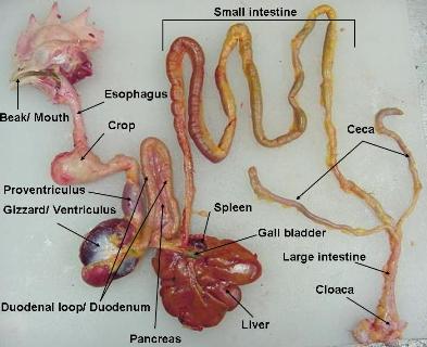
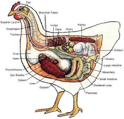
The chicken has a typical avian digestive system. In chickens, the digestive tract (also referred to as the gastrointestinal tract or GI tract) begins at the mouth, includes several important organs, and ends at the cloaca. Figure 1 shows a chicken digestive tract, and Figure 2 shows the location of the digestive tract in the chicken’s body.
Beak/Mouth
As with most birds, a chicken obtains feed by using its beak. Food picked up by the beak enters the mouth. Chickens do not have teeth, so they cannot chew their food. However, the mouth contains glands that secrete saliva, which wets the feed to make it easier to swallow. Also, the saliva contains enzymes, such as amylase, that start the digestion process. The chicken uses its tongue to push the feed to the back of the mouth to be swallowed.
Esophagus
The esophagus is a flexible tube that connects the mouth with the rest of the digestive tract. It carries food from the mouth to the crop and from the crop to the proventriculus.
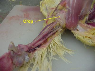
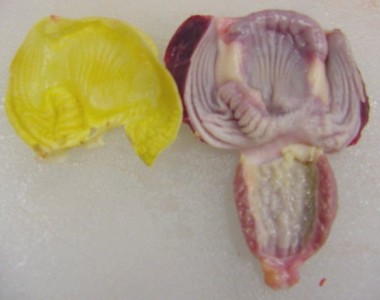
Crop
The crop is an out-pocketing of the esophagus and is located just outside the body cavity in the neck region (see Figure 3). Swallowed feed and water are stored in the crop until they are passed to the rest of the digestive tract. When the crop is empty or nearly empty, it sends hunger signals to the brain so that the chicken will eat more.
Although the digestive enzymes secreted in the mouth began the digestion process, very little digestion takes place in the crop—it is simply a temporary storage pouch. The crop evolved for birds that are typically hunted by other animals but need to move to the open to find feed. These birds can consume relatively large amounts of food quickly and then move to a more secure location to digest that food.
Occasionally, the crop becomes impacted, or backed up. This problem—called crop impaction, crop binding, or pendulous crop—can occur when a chicken goes a long time without feed and then eats too much too quickly when feed is available again. Crop impaction also can occur when a chicken free ranges on a pasture of tough, fibrous vegetation or eats long pieces of string. With crop impaction, even if a chicken continues to eat, the feed cannot pass the impacted crop. The swollen crop also can block the windpipe, causing the chicken to suffocate.
Proventriculus
The esophagus continues past the crop, connecting the crop to the proventriculus. The proventriculus (also known as the true stomach) is the glandular stomach where digestion primarily begins. Hydrochloric acid and digestive enzymes, such as pepsin, are added to the feed here and begin to break it down more significantly than the enzymes secreted by the salivary glands. At this point, however, the food has not yet been ground—this organ is called the proventriculus because its location in the digestive tract is before the ventriculus, where food is ground (see Figure 4).

Ventriculus (Gizzard)
The ventriculus, or gizzard, is a part of the digestive tract of birds, reptiles, earthworms, and fish. Often referred to as the mechanical stomach, the gizzard is made up of two sets of strong muscles that act as the bird’s teeth and has a thick lining that protects those muscles (see Figure 5). Consumed feed and the digestive juices from the salivary glands and proventriculus pass into the gizzard for grinding, mixing, and mashing.
When allowed to free-range, chickens typically eat small stones. The acidic environment in the proventriculus softens the stones, and then the strong muscles of the gizzard grind them into tiny pieces. The stones remain in the gizzard until they are ground into pieces small enough to pass to the rest of the digestive tract.
Grit, a commercial product made up of small stones, can be used as a supplement to chicken feed. Chickens fed only commercially prepared feed do not need grit. Chickens that eat whole grains or chickens kept on pasture that do not consume enough pebbles with the forage typically require a supplementation of grit. Grit should not be confused with limestone or oystershell, which are given to laying hens as sources of calcium for their eggs’ shells.
When a chicken eats a small, sharp object, such as a tack or staple, the object is likely to get stuck in the gizzard. Because of the strong grinding motion of the gizzard’s muscles, such sharp objects can put holes in the gizzard wall. Chickens with damaged gizzards grow thin and eventually die. Preventing this situation is a good reason to keep a poultry house free of nails, glass shards, bits of wire, and so on.
Small Intestine
The small intestine is made up of the duodenum (also referred to as the duodenal loop) and the lower small intestine. The remainder of the digestion occurs in the duodenum, and the released nutrients are absorbed mainly in the lower small intestine.
The duodenum receives digestive enzymes and bicarbonate (to counter the hydrochloric acid from the proventriculus) from the pancreas and bile from the liver (via the gall bladder). The digestive juices produced by the pancreas are involved primarily in protein digestion. Bile is a detergent that is important in the digestion of lipids and the absorption of fat-soluble vitamins (A, D, E, and K).
The lower small intestine is composed of two parts, the jejunum and the ileum. The Meckel’s diverticulum marks the end of the jejunum and the start of the ileum (see Figure 6). The Meckel’s diverticulum is formed during a chicken’s embryonic stage. In the egg, the yolk sac supplies the nutrients needed for the embryo to develop and grow. Right before hatch, the yolk sac is taken into the navel cavity of the embryo. The residual tiny sac is the Meckel’s diverticulum.

Ceca
The ceca (plural form of cecum) are two blind pouches located where the small and large intestines join. Some of the water remaining in the digested material is reabsorbed here. Another important function of the ceca is the fermentation of any remaining coarse materials. During this fermentation, the ceca produce several fatty acids as well as the eight B vitamins (thiamine, riboflavin, niacin, pantothenic acid, pyridoxine, biotin, folic acid, and vitamin B12). Because the ceca are located so close to the end of the digestive tract, however, few of the produced nutrients are absorbed and available to the chicken.
Large Intestine (Colon)
Despite the name, the large intestine is actually shorter than the small intestine. The large intestine is where the last of the water reabsorption occurs.
Cloaca
In the cloaca, the digestive wastes mix with wastes from the urinary system (urates). Chickens usually void fecal material as digestive waste with uric acid crystals on the outer surface—that is, chickens do not urinate. The color and texture of chicken fecal material can indicate the health status of the chicken’s digestive tract: the white, pasty material coating chicken fecal material is uric acid, the avian form of urine, and is normal.
The reproductive tract also exits through this area. When a hen lays an egg, the vagina folds over to allow the egg to leave through the cloaca opening without coming into contact with feces or urine.
Intestinal Microflora
Both the small and large intestines normally are populated with beneficial organisms (bacteria, yeast, etc.), referred to as microflora (micro meaning “small” and flora meaning “plants”). This microflora aid in digestion.
When chicks hatch, their digestive tracts are virtually sterile. If raised by a mother hen, a chick obtains the beneficial microflora by consuming some of its mother’s fecal material. In artificial incubation and brooding, chicks do not have this option. In such situations, producers can provide the chicks with probiotics, which are preparations containing the beneficial microflora that normally inhabit a chicken’s digestive tract. Through the probiotics, the chicks receive the beneficial bacteria they need to fight off infection by pathogenic bacteria, such as salmonella.
Intestinal disease in chickens normally occurs when the balance of normal microflora is upset—that is, the normal microflora is overrun by too many foreign organisms. The result is enteritis or inflammation of the intestines. Enteritis produces symptoms that include diarrhea, increased thirst, dehydration, loss of appetite, weakness, and weight loss or slow growth. Severe damage to the intestinal tract typically is called necrotic enteritis (necrotic meaning “dead tissue”), which is a problem in many types of production systems.